Main characteristics
- Confocal Raman imaging with unprecedented high speed, high sensitivity and high resolution
- A complete Raman spectrum is acquired for each sample point in the hyperspectral image.
- Excellent horizontal resolution
- Excellent longitudinal resolution, ideal for 3D imaging and depth analysis
- Ultra-fast Raman imaging with single-spectrum integration time below 1 ms
- Spectral acquisition with ultra-high flux lenses for highest sensitivity and best spectral resolution
- Non-destructive imaging technology: no need to stain or label samples
Examples of applications

Red: compressive strain, blue: tensile strain.


Scanning points: 150 × 150 × 23 = 517,500 Raman spectra, scanning area: 30 μm × 30 μm × 11.5 μm, total acquisition time: 23 minutes.
technical specification
Raman routine mode of operation
- Raman spectral imaging: one complete Raman spectrum is acquired for each sample point
- Manual positioning of samples for scanning in the plane (x-y direction) and longitudinal direction (z direction)
- Image Overlay: 3D Confocal Raman Imaging
- time series (stats.)
- Single-point Raman spectral acquisition
- Single-point depth sequence acquisition
- Fiber-coupled Ultra-High-Throughput Spectrometer (UHTS), designed for low-light Raman microscopy
- confocal fluorescence microscope
- open-field microscopy
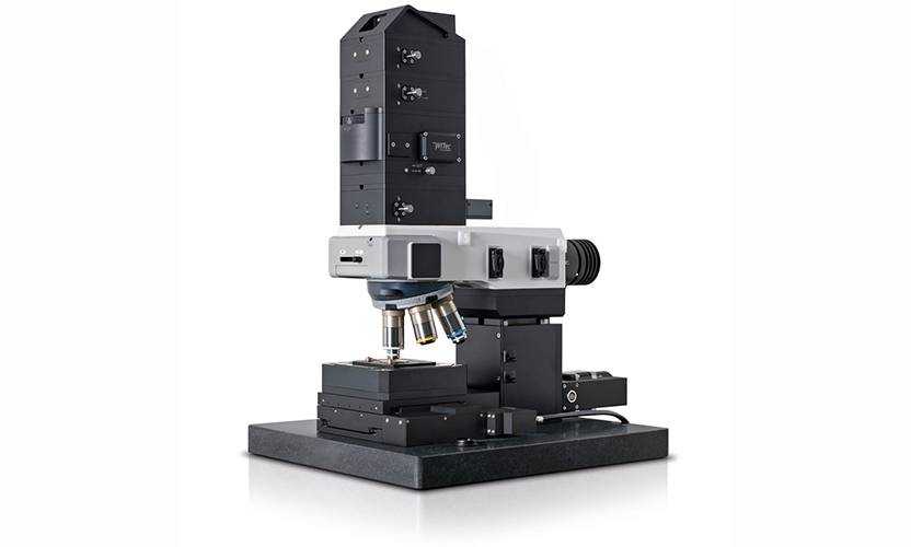
fundamental characteristic
- Research grade 6-port objective rotating wheel optical microscope
- Camera System: CCD Camera
- LED White Light Source Kohler Lighting
- Manual sample positioning in x-y direction
- fiber optic coupling
Raman selectable/upgradeable operating modes
- Various excitation wavelengths can be selected
- A wide range of ultra-high-throughput spectrometers are available (UV, VIS, NIR)
- Automated motorized sample positioning and spectra collection with 25 mm panning range (50 mm optional)
- Automated Confocal Raman Imaging
- Automatic multi-area, multi-point measurement
- Fully automated: see details alpha300 apyron
- Optional ultra fast Raman imaging
- Scalable Drop Shot Fluorescence Applications
- Configurable adapters for higher samples
- TrueSurface In-depth analysis
- autofocus
- Options: Dark Field Microscope, Phase Difference Microscope and Differential Interference (DIC) Microscope
Ultra High Throughput Spectrometer UHTS
- Optimized spectrometers (UV, VIS or NIR) based on various lenses and excitation wavelengths designed for Raman microscopy and weak signal applications
- Fiber-coupled ultra-high throughput optical system
- Spectral peaks are symmetrical and free of aberration
software interface
- outfit with WITec Professional SoftwareFor instrument control, data acquisition and data processing.