
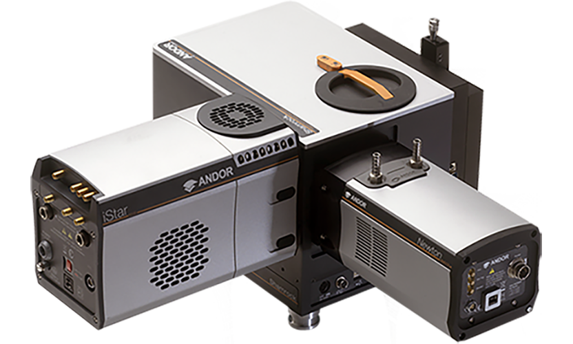
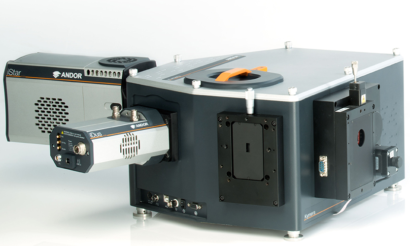
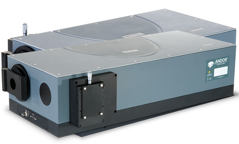
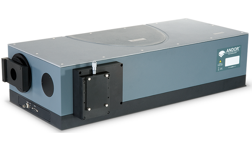
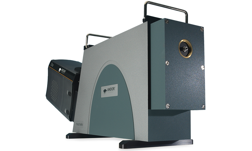
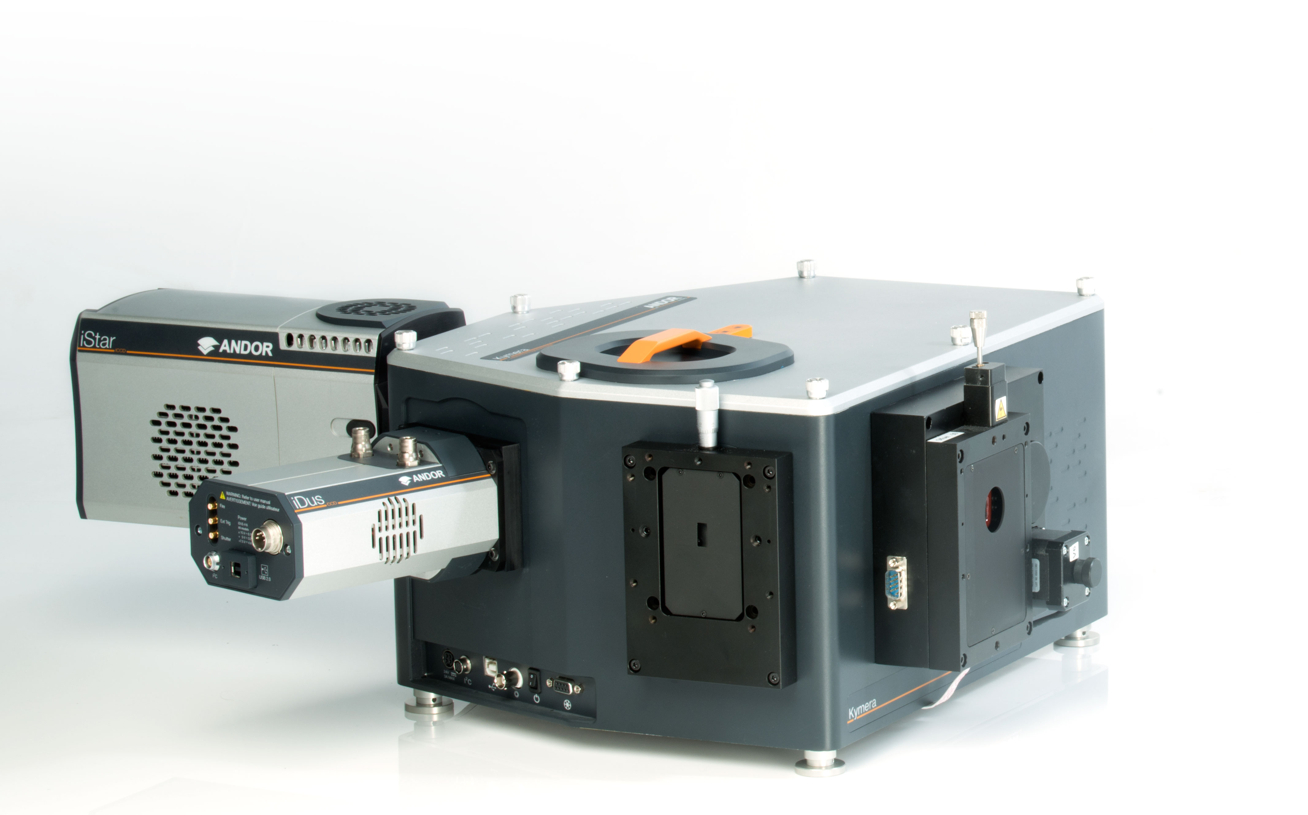
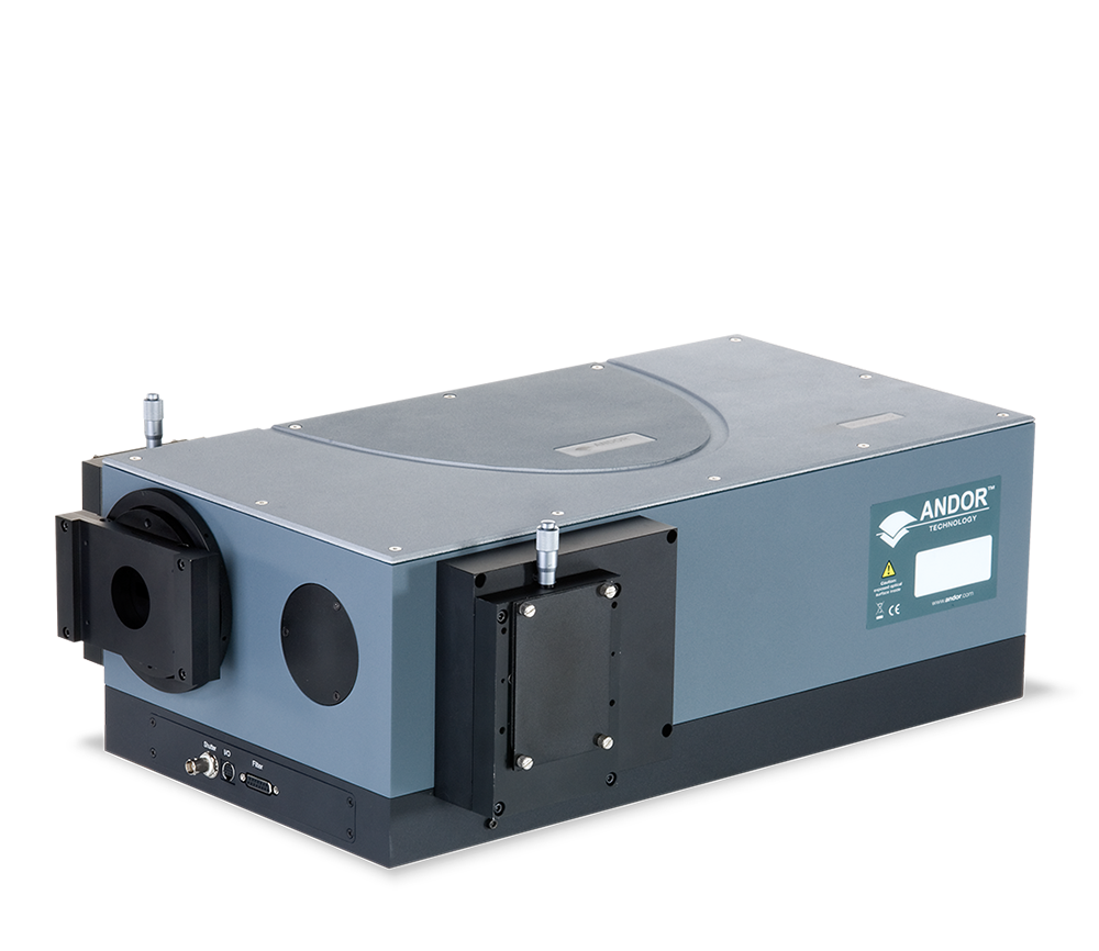
| model number | Kymera 193i | Kymera 328i | Shamrock 500i | Shamrock 750i | Mechelle 5000 |
| photograph | 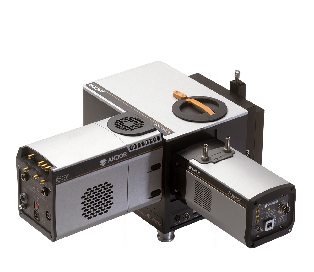 |
 |
 |
 |
 |
| relative aperture | F/3.6 | F/4.1 | F/6.5 | F/9.8 | F/7 |
| anxious | 193 mm | 328 mm | 500 mm | 750 mm | 195 |
| resolution (of a photo) | 0.21nm | 0.1 nm | 0.06 nm | 0.04 nm | 0.01 nm |
| bandwidths | 98 nm | 61 nm | 40 nm | 28 nm | 750 nm |
| encoder | Two-raster | quadrangle | Triple Grating | Triple Grating | Medium Step Grating |
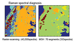
Raman: MSH diagnostics yielded only 350 spectra, while raster scanning yielded 40,000 |
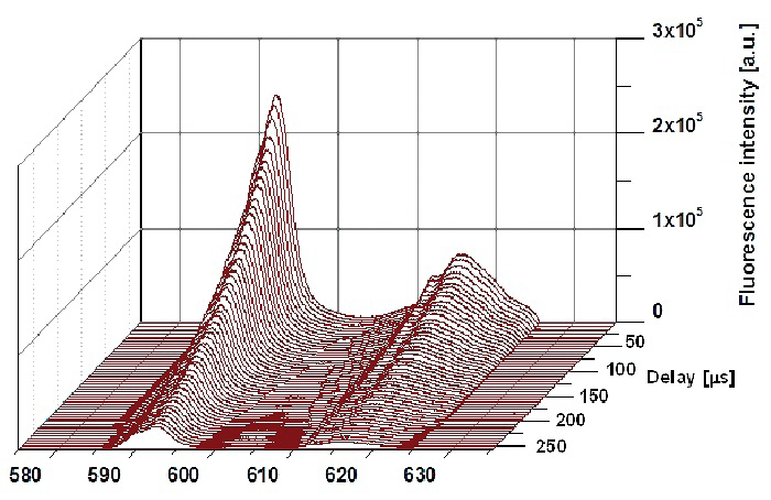
Luminescence: Eu(III) Luminescence at Different Delay Times |
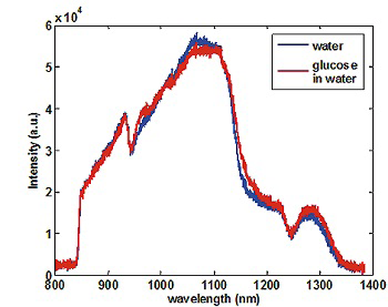
Absorption: Transmission data for background-corrected water and high-concentration glucose solution at an exposure time of 10 ms |
 LIBS: Chalcopyrite rock cuttings surface lead content chemistry: cuttings on the left, lead content chemistry on the right LIBS: Chalcopyrite rock cuttings surface lead content chemistry: cuttings on the left, lead content chemistry on the right |

Microspectroscopy: Comparison of images obtained from tissue regions showing, left) histopathological image obtained after Raman analysis, right) 2D pseudo-color image obtained from Raman measurements performed in vitro. Color scale:dermis yellow, epidermis cyan, basal cell carcinoma (BCC) dark blue |
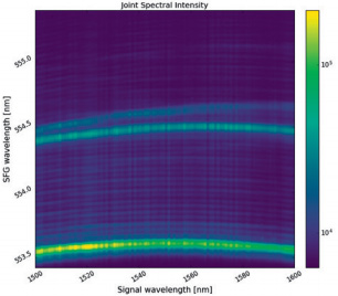
Nonlinear spectra: sum-frequency spectral intensities of SFG processes |
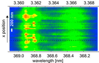 Materials Science : Spatially resolved spectra of ZnO thin films at 2.8 μm reveal DX resonance fine structure Materials Science : Spatially resolved spectra of ZnO thin films at 2.8 μm reveal DX resonance fine structure |
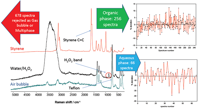 Chemical processes: spectra of the organic (styrene) phase (red), the aqueous phase (black), and where bubbles occupy the focal volume of the probe. These spectra were extracted from the 1000 spectra obtained Chemical processes: spectra of the organic (styrene) phase (red), the aqueous phase (black), and where bubbles occupy the focal volume of the probe. These spectra were extracted from the 1000 spectra obtained |
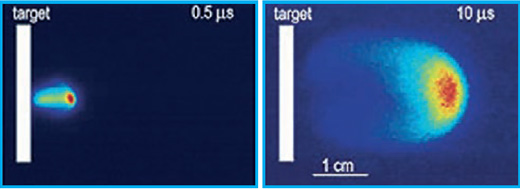 PLIF: LIF images of plasma plume expansion taken with an ICCD camera at two different delay times of 0.5µs and 10µs PLIF: LIF images of plasma plume expansion taken with an ICCD camera at two different delay times of 0.5µs and 10µs |
